-
- Cedars-Sinai Medical Centre has made a scientific advancement relating to Alzheimer’s disease.
- Their research deduced that retinal image studies could be used for detecting early signs of Alzheimer’s
- The formation of beta-amyloid that is a biomarker of neurodegenerative disease is commonly found in retinal tissues.
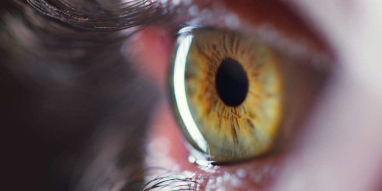
THD NewsDesk, Washington: Scientists of Cedars-Sinai Medical Center have made breakthrough progress in medical science by discovering a unique method of identifying early signs of Alzheimer’s. A recent study conducted by the experts found how the early onset of Alzheimer’s could be spotted and diagnosed accordingly through retinal images.
The study, Alzheimer’s & Dementia: Diagnosis, Assessment & Disease Monitoring, required the participants to undergo sectoral retinal amyloid imaging. The retinal images are then used to locate specks of retina amyloid protein that could sign Alzheimer’s.
Alzheimer’s disease is found to cause cognitive deterioration due to a build-up of beta-amyloid in the brain. The study suggests that this component was usually detected in retinal tissues.
Co-author Maya Koronyo-Hamaoui said the following in a news release,
“We also suggested a particular type of immune-modulation therapy that may combat the disease by reducing toxic proteins and harmful inflammation in the brain and, in return, enhancing a protective type of immune response that preserved the connections between neurons, which are tightly connected to cognition.”
Scientists believe that the remarkable finding on the neurodegenerative illness is promising. Moreover, retinal imaging studies should be used as a potential biomarker for early symptoms of Alzheimer’s disease.
Source: ANI
US scientists discover unique way of spotting Alzheimer’s through Retinal images
A new study conducted by experts from Cedars-Sinai says retina amyloid protein could indicate early signs of Alzheimer's
0
0
SHARES
About Us
A daily dose of news, views, opinions, and actionable information from the global healthcare industry. Our newsletter is available for a limited audience. If you are interested to write or contribute to this portal then please get in touch.


